Research Section 1: Craniosynostosis
Craniosynostosis is a congenital condition where the early fusion of cranial sutures affects skull shape, potentially impacting brain growth and neurological development. The condition varies from single to complex multisuture synostosis, each requiring customized treatment to achieve a normal head shape. This is crucial for both aesthetics and ensuring adequate cranial volume to support brain growth and minimize developmental risks. Our research focuses on overcoming these challenges to achieve outcomes that closely match normal cranial anatomy.
Understanding Craniofacial Growth and Asymmetry
Achieving a head shape that conforms to normative standards is essential in craniofacial surgery for both aesthetic and functional reasons. We conduct annual assessments of healthy students over seven years to create a comprehensive database of craniofacial measurements. This data helps us understand normal growth patterns and asymmetry, providing a strong foundation for future research and clinical practices in craniofacial surgery.

Innovations in Craniosynostosis Surgery
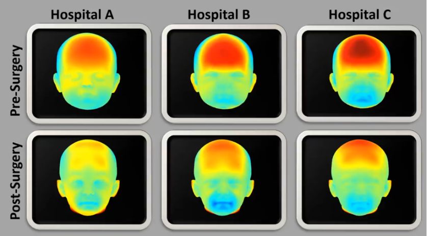
Recent surgical advancements, like endoscopic strip craniectomy, offer minimally invasive options with fewer complications. Our multicenter 3D imaging study, in collaboration with leading medical centers, compares pre- and post-surgery outcomes to ensure the most effective treatment methods.
Diagnosing Craniosynostosis with AI
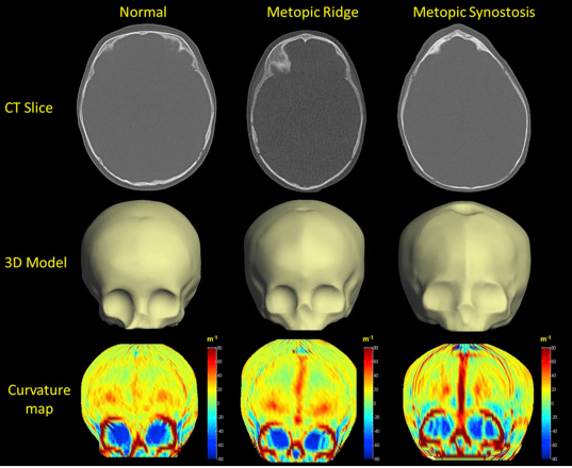
Differentiating between metopic craniosynostosis, which often requires surgery, and a benign metopic ridge can be challenging. We are developing AI tools to provide a reliable, non-invasive method for accurately identifying these conditions, improving clinical decision-making and patient outcomes.
Research Section 2: Cleft Lip and Palate
Cleft lip and palate are among the most common congenital facial anomalies, affecting both appearance and facial function, including speech, eating, and breathing. Our research focuses on understanding and addressing the dynamic asymmetry caused by these conditions, with the goal of enhancing surgical outcomes and improving the quality of life for affected individuals.
Enhancing Facial Reconstruction with Dynamic Symmetry Analysis
Our center leads in using 4D motion capture technology to analyze facial expressions in real-time, providing critical benchmarks for surgical interventions. This method helps us understand facial dynamics, which is essential for achieving natural facial movements post-surgery, thereby improving patients' quality of life by restoring their facial expressions to a near-natural state.
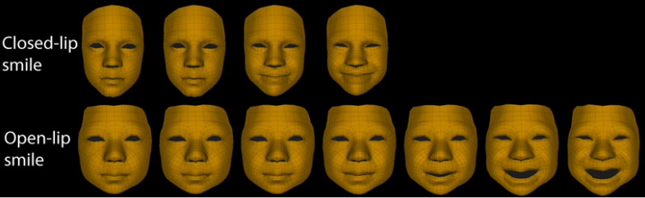
Assessing Surgical Outcomes in Cleft Lip Patients
Through dynamic image analysis, we capture and evaluate facial expressions post-surgery to assess the restoration of natural facial movements. This approach offers insights beyond static results, helping us measure the effectiveness of cleft lip repairs.

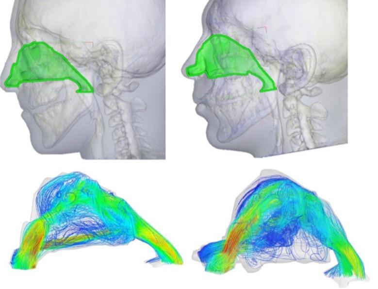
Assessing Nasal Breathing Improvements
Our research also focuses on improving nasal breathing and maxillary growth in Cleft Lip and Palate (CLP) patients. We use advanced Computational Fluid Dynamics and patient-specific CT scans to analyze the effectiveness of the LeFort I osteotomy, a key surgical procedure. This approach allows for a detailed assessment of nasal airflow, enhancing both the functional and aesthetic outcomes of CLP treatments.
Research Section 3: Congenital Ear Deformity
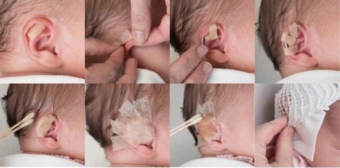
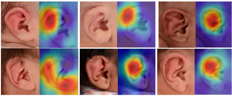
Early detection of congenital ear deformities is vital for successful non-surgical treatment, taking advantage of the natural flexibility of a newborn's auricular cartilage, which is influenced by maternal estrogens. Since this flexibility diminishes within the first six weeks after birth, prompt diagnosis is essential. Our research is focused on developing an artificial intelligence (AI) tool to enhance early and objective detection of ear deformities, potentially revolutionizing the assessment and treatment of these conditions in infants.
Research Section 4: Osteosarcoma
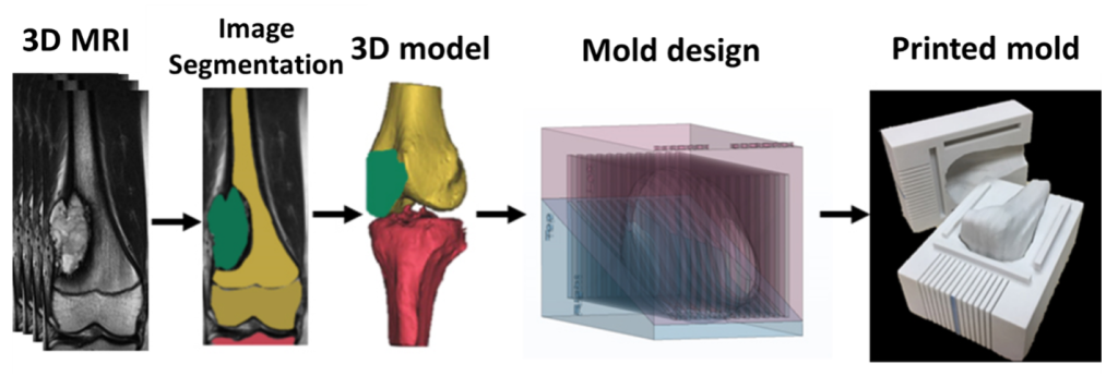
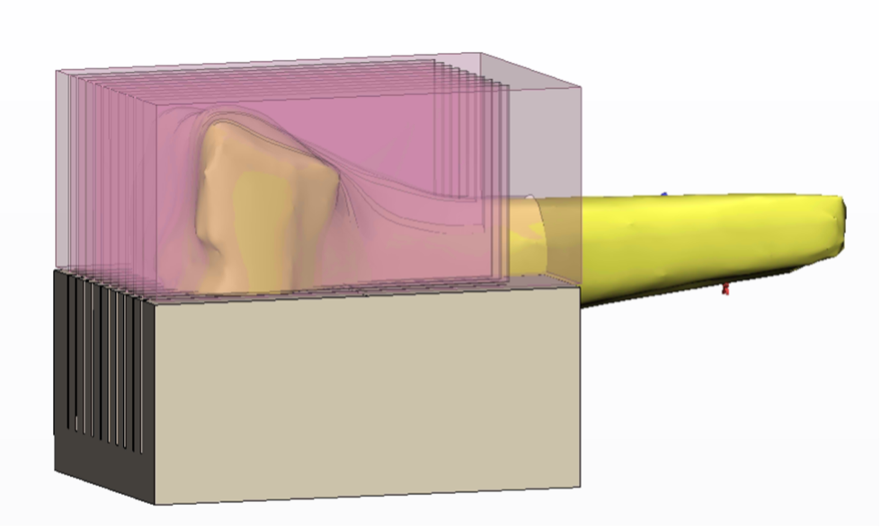
Osteosarcoma, the most common bone cancer in children and adolescents, demands precise diagnosis and treatment for effective management. Traditional methods have struggled to accurately correlate radiological images with pathological findings. Our research introduces a groundbreaking method using 3D printing technology to enhance the diagnosis and monitoring of osteosarcoma treatments.
Osteosarcoma Diagnosis and Treatment Using 3D Printing
We utilize custom-designed molds created from MRI scans of the affected bone to precisely cut bone specimens for pathological examination. This innovative approach ensures a better match between MRI images and actual tumor sections, leading to more accurate diagnoses and a deeper understanding of tumor characteristics.