Facilities
Most clinical and translational research activities are performed at the UT Southwestern William P. Clements Jr. University Hospital and the UT Southwestern Outpatient Building.
Facilities include patient waiting and changing areas, nursing pre-procedure and recovery areas, and large, comfortable scanning rooms.
Equipment
Clinical equipment includes EPIQ Elite, LOGIQ E10, and New ACUSON Sequoia ultrasound devices with a variety of 2D and 3D clinical transducers (see details below), Trophon high-level probe disinfection, and image fusion and navigation systems.
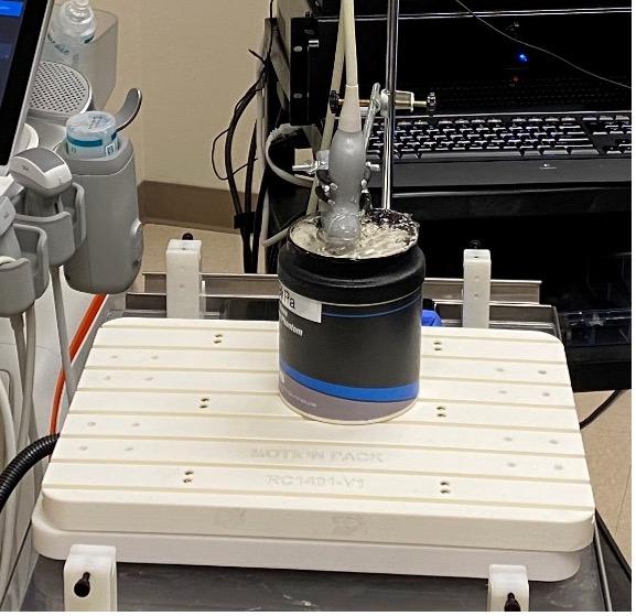
Research equipment includes programmable motion platform for phantom scanning, Verasonics Vantage 256 programmable ultrasound system equipped with L11-4v and L22-14v transducers, and clinically-equivalent research devices for micro-Doppler, shear wave elastography, attenuation imaging, Ultrasound-Derived Fat Fraction, and contrast-enhanced ultrasound with post-processing including perfusion analysis.
Phantoms include multi-modality ultrasound + CT abdominal phantom, pelvic 3D phantom, shear wave elasticity phantom, and various small parts phantoms.
| C1-6 | C2-9 | L2-9 | L3-12 | L6-24 | ML6-15 | ML4-20 | RIC 5-9-D |
| MVI (MicroVascular Imaging) | PDI (Power Doppler Imaging) | B - FLOW | Contrast | Elasto - Strain | Ealasto - Shear Wave | UGAP | 3D | RF Capture Tool |
| C5-1 | C2-9 | mC7-2 | L12-3 | L12-5 | L18-5 (old) | eL18-4 (new) | L17-7io (hockey) | XL14-3 | X6-1 | C10-3v | 3D9-3v |
| CPA (Color Power Angio) | MFI (MicroFlow Imaging) | MFI HD | Contrast | ELASTO (Strain) | ELASTO (PQ/EQI) | ATTEN | HRI (Hepato-Renal Index) | IQ Capture Tool |
| 5C1 | 9C3 | DAX | 11M3 | 4V1 | 7L4 | 10L4 | 14L5 | 15L4 | 18L6 | 18H6 | 9VE4 |
| POWER (Doppler) | SLOW FLOW | Contrast | PSWE (Point Shear Wave) | (2D) SWE | AUTO PSWE | UDFF | 3D | RF Capture Tool |